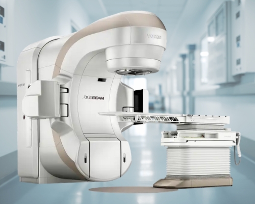
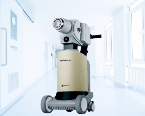
Varian HDR Brachytherapy Machine
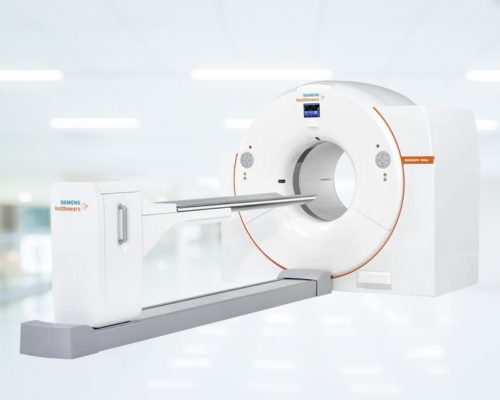
Mammography is an X-ray imaging technique designed to capture detailed images of the breast. During the procedure, the breast is briefly compressed between two thin plates while exposed to low doses of radiation. This compression allows for clearer imaging. Mammography is crucial for the early identification of breast cancer, as it can reveal distinctive masses or tiny calcium deposits known as microcalcifications. At Sharda Care - Healthcity, the mammography machine provides advanced imaging capabilities, offering both 2D and 3D views. This enhanced imaging technology facilitates more accurate and earlier detection of potential issues, improving diagnostic precision.
How Does It Help?
Mammography technology plays a crucial role in both diagnosing and screening for breast abnormalities. It is typically employed to evaluate breast lumps, providing detailed images that help differentiate between benign and malignant conditions.
Diagnostic Mammography
It is employed to investigate unusual changes in the breast, including lumps, persistent pain, abnormal skin textures, nipple thickening, or discharge. A diagnostic mammogram involves taking extra images to provide a clearer view of these suspicious areas. This technique is also utilized for comprehensive evaluation, offering detailed insights into breast health and aiding in accurate diagnosis.
Screening Mammography
For screening purposes, it is used to detect early signs of breast cancer in individuals who may not exhibit symptoms, allowing for intervention before the disease progresses. This proactive approach significantly enhances the likelihood of successful treatment and improves overall breast health outcomes.
How Is It Done?
Preparation
- Schedule your mammogram 1 to 2 weeks after your menstrual period to reduce discomfort.
- If your breasts are tender, it’s best to postpone the test.
- You will be given a robe at the hospital to cover your upper body.
- Inform the radiologist about any changes in breast lumps since your last examination.
- If you have breast implants, make sure to let your doctor know in advance.
Treatment
During a mammography procedure, the patient is given an open-front gown to wear from the waist up, provided by the mammography staff. A technologist will guide the patient into the correct position in front of the machine and carefully place each breast, one at a time, onto the equipment. Proper positioning of the body and breasts is essential for capturing the most accurate images. To ensure the breast tissue is adequately spread, it is gently compressed between two flat plates. Once compression and positioning are correct, the radiologist will either step behind a protective screen or briefly leave the room. After confirming everything is in place, the radiologist activates the X-ray source, which captures images of the breast tissue on X-ray film or a digital detector. The entire process usually lasts about 10 to 15 minutes.
Post Treatment
What happens after Mammography?
After mammography, the images are saved as electronic data files and are typically reviewed on a computer screen. At Sharda Care - Healthcity, a radiologist who specializes in women’s imaging carefully examines these images and prepares the results. The report is then sent to your doctor and is available in the form of a film or a CD, which can be picked up when collecting the report.
What Are The Benefits and Risks Of Mammography?
Benefits
Mammography plays a crucial role in the early detection of breast cancer, allowing doctors to identify abnormalities even before symptoms appear. This early detection helps doctors determine the most effective treatment plan and gives patients valuable time to address the condition proactively. Beyond just spotting cancer, mammography can also detect other breast changes such as lumps or microcalcifications, enhancing overall breast health monitoring. The procedure significantly improves the chances of successful treatment, reduces the need for aggressive therapies, and can lead to better outcomes and survival rates.
Risks
- Exposure to low-dose radiation: While mammograms involve low levels of radiation, the exposure is minimal and generally safe, with the benefits far outweighing the potential risks.
- Results may not always be precise: The accuracy of mammogram results depends significantly on the radiologist’s skills and the techniques used, and can also be affected by factors like age and breast density.
- Challenges in younger women: High breast density in younger women can make mammograms more difficult to interpret, increasing the chance of inaccurate results.
- Potential for additional tests: Approximately 10% of mammograms may lead to further testing for clearer evaluation.
- Limitations in cancer detection: Mammograms may miss very small tumors or those located in challenging areas, such as near the armpits, making it difficult to detect all cancers.
What Makes Mammography Different?
- Mammography is a specialized imaging technique that uses low-dose X-rays to detect breast cancer at its earliest stages, often before any symptoms appear.
- Sharda Care - Healthcity utilizes advanced technology with the Siemens Mammomat, powered by PRIME technology, which provides the benefit of up to 30% lower radiation exposure without compromising image quality.
- The system features a direct-to-digital aSe detector that enables radiologists to capture high-quality images quickly and efficiently.
Varian HDR Brachytherapy Machine
The Varian HDR Brachytherapy Machine is an advanced medical device used in cancer treatment. It delivers high doses of radiation directly to tumors in short, precise sessions. This computer-controlled machine carefully places tiny radioactive sources inside or next to cancerous growths, allowing doctors to target the disease while minimizing harm to nearby healthy tissue. It's versatile enough to treat various cancer types, including prostate, breast, lung, and cervical cancers. The machine works by guiding the radioactive material through thin tubes to the tumor site for a brief period, then quickly removing it. This approach enables powerful, focused treatments that typically last only minutes but may be repeated several times for maximum effectiveness. By combining accurate planning, precise delivery, and quick treatment times, the Varian HDR Brachytherapy Machine represents a significant tool in modern cancer therapy.
How Does It Help?
Brachytherapy radiations are enclosed in a protective capsule or wire. It allows the ionizing radiation to kill the surrounding tissues. Varian HDR Brachytherapy Machine
It stops the cancerous cell to grow and divide into intervals.
Prostate Cancer
The Varian HDR Brachytherapy Machine is a powerful tool for treating prostate cancer. It works by precisely delivering high doses of radiation directly to the prostate gland. This targeted approach helps kill cancer cells while reducing harm to nearby healthy tissues. The treatment is quick, with each session lasting only about 15 minutes. Patients often need fewer treatments compared to other radiation methods. The machine's computer-controlled system allows doctors to tailor the radiation to fit each patient's unique needs. This can lead to better results and fewer side effects. For many men with prostate cancer, this treatment offers an effective option that can be completed with minimal disruption to their daily lives.
Cervix Cancer
The Varian HDR Brachytherapy Machine offers a precise treatment option for cervical cancer. It works by inserting a thin applicator into the cervix, through which tiny radioactive sources can be guided. These sources deliver high doses of radiation directly to the tumor, sparing healthy tissues nearby. The treatment is typically quick, lasting only a few minutes per session, and can be completed over several visits. This approach allows for intense, targeted therapy that can effectively shrink tumors and kill cancer cells. The machine's computer-controlled system ensures accurate placement and timing of radiation delivery. For many women with cervical cancer, this treatment provides a powerful tool to fight the disease while minimizing side effects and hospital stays.
How Is It Done?
Preparation
Preparing for Brachytherapy
- The doctor will guide the patient on specific preparations required before brachytherapy, which can vary significantly depending on the patient’s condition. Typically, these preparations involve avoiding certain medications and undergoing diagnostic tests like MRI or CT scans.
- Following an initial assessment, the radiation oncologist determines the most suitable therapy for the patient. The oncologist carefully evaluates the exact location and size of the target area to decide the precise amount of radiation needed for effective treatment.
Procedure
- During the procedure, the patient will be given mild anesthesia to numb the specific area where the incision will occur.
- A needle containing radioactive seeds is then carefully inserted directly into the tumor. Once the seeds are in place, the needle or device is withdrawn, leaving the radioactive seeds embedded within the tumor.
- To ensure the seeds are positioned correctly, additional imaging tests like an MRI or CT scan may be performed.
After Treatment
- After the procedure, the physician will provide detailed post-treatment care instructions, and the patient can return home once the therapy is finished.
- Mild swelling or tenderness at the treatment site may occur, but most patients who undergo Brachytherapy can typically resume their normal activities within a few weeks to months.
- Regular follow-up appointments with the physician are crucial to monitor the patient’s progress and assess whether the condition remains stable or shows any changes. These visits also allow the patient to discuss any side effects they may be experiencing.
What Are The Benefits and Risks Of Varian HDR Brachytherapy Machine?
Benefit: The Varian HDR Brachytherapy machine delivers highly targeted radiation directly to the tumor, minimizing exposure to surrounding healthy tissues and reducing overall treatment time compared to traditional radiation therapies.
Risk: There is a potential for localized side effects, such as skin irritation, pain, or swelling at the treatment site, and in rare cases, there may be complications related to incorrect dose delivery.
What Makes Varian HDR Brachytherapy Machine So Unique?
The Varian HDR Brachytherapy machine is unique due to its precision and efficiency in cancer treatment. It delivers high-dose radiation directly to the tumor with pinpoint accuracy, sparing surrounding healthy tissues. The machine's advanced imaging and software allow for customized treatment planning, ensuring optimal dose distribution tailored to each patient's anatomy. Its flexibility to treat various cancer types, including prostate, cervical, and breast, enhances its versatility. Additionally, Varian HDR Brachytherapy reduces overall treatment time compared to traditional methods, offering quicker, more effective sessions with improved patient outcomes and minimized side effects, making it a standout option in modern radiation therapy.
Looking for an Expert
Sharda Care - Healthcity is home to some of the eminent Doctors in the world.
Book an Appointment